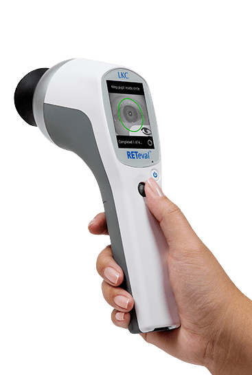
Electroretinography (ERG) - RETeval®
A powerful aid in the diagnosis and management of retina and optic nerve diseases such as diabetic retinopathy and glaucoma.
What is electroretinography (ERG)?
Electroretinography is an electrophysiological test of the retina, the layer of the eye which detects light. The electroretinogram (ERG) is to the retina what the electrocardiogram (ECG) is to the heart. Just as an ECG is crucial to diagnosing illness and monitoring the heart’s function, ERG plays a critical role in the care of the eye, and is instrumental in the early detection of retinal dysfunction.
What is an Electroretinogram?
The electroretinogram (ERG) is an examination that evaluates the function of the eye’s retinal cells. Patients’ eyes are stimulated with light and the resulting electrical activity from their retinal cells is measured by skin or corneal electrodes. Dependent on the type, intensity and colour of the light stimulus information on different areas and types of cells can be obtained.
Diagnosis and Tracking of Glaucoma
The RETeval® PhNR testing is a vital tool in the management of patients with Glaucoma and other optic nerve diseases with objective ERG assessment of ganglion health. Early diagnosis is essential to preserve vision loss from further degeneration of ganglion cells.
The RETeval® PhNR test objectively measures the ganglion cells function by evaluating the electrical activity of the cells to a light stimulus. Knowing about the function of the ganglion cells assist in the detection and tracking of glaucomateous changes in any patient.
Diagnosis and Tracking of Diabetic Retinopathy
RETeval ® is an objective tool to measure diabetic retinopathy progression risk like no other. The rapid and non-invasive RETeval ® device combines retina cell stress with pupil light response. This powerful combination provides a superior progression risk assessment.
How It Works
RETeval device will start flashing light into the patient’s eye.
The retina responds to the flashes by generating small electrical signals that travel through the facial structure to the sensor strip.
The Sensor Strip detects the electrical signals and compares the results to the age-adjusted reference database.